18318 University Blvd #500, Sugar Land, TX 77479
Advanced 3D Dental Imaging
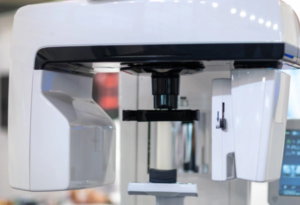
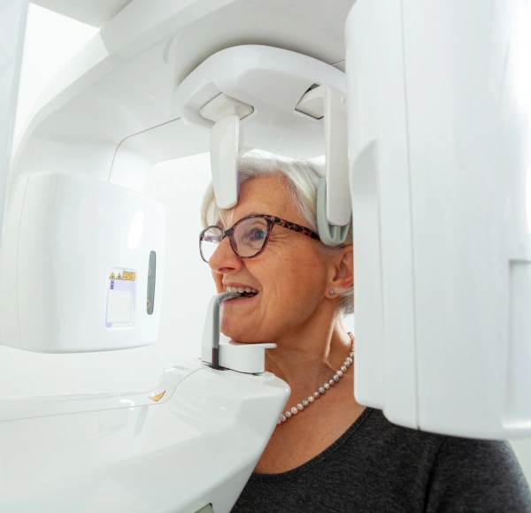
Conventional X-rays produce flat, two-dimensional images that show only small sections of the mouth. In contrast, 3D Cone Beam CT (CBCT) delivers a comprehensive, three-dimensional view of the teeth, jaw, sinuses, and bone structures—enabling highly accurate diagnosis and treatment planning from all perspectives.
How 3D CT Imaging Works
Traditional dental X-rays produce flat, two-dimensional images of small areas, offering only a limited look at specific parts of the mouth. 3D CT imaging—also known as Cone Beam Computed Tomography (CBCT)—captures a detailed, three-dimensional view of the entire oral and facial structure.
Think of the difference between looking at a single photograph of a tooth versus being able to rotate a complete 3D digital model. CT imaging gives us that level of clarity. It reveals not only the teeth, but also the surrounding jawbone, sinuses, and other anatomical features. This advanced technology allows for more accurate diagnosis, more precise treatment planning, and care that is truly tailored to your individual needs.
Why We Utilize 3D CBCT Imaging
CBCT imaging delivers a complete, detailed view that supports safer, more precise, and more predictable dental care. This technology is especially valuable for:
- Evaluating and developing treatment plans for TMJ (temporomandibular joint) concerns
- Designing customized surgical guides for highly accurate implant placement
- Planning procedures such as extractions and other oral surgeries with greater precision
- Reviewing existing root canal treatments and uncovering infections that may not appear on 2D X-rays
- Measuring airway space and detecting possible obstructions linked to sleep apnea
- Revealing infection, bone changes, or complications at previous extraction sites
The Difference 3D CT Makes in Modern Dentistry
3D CBCT imaging offers exceptional accuracy, visibility, and safety—allowing every treatment to be thoughtfully planned and personalized to your individual needs.
Visual clarity
Patients can view high-resolution 3D images of their teeth and jaws directly on screen, making it easier to understand concerns and recommended treatments.
Enhanced Communication
Dentists can enlarge specific areas, point out findings, and explain conditions more clearly and confidently.
Streamlined Coordination
Digital scans can be quickly shared with labs, specialists, or insurance providers, helping expedite approvals and treatment timelines.