18318 University Blvd #500, Sugar Land, TX 77479
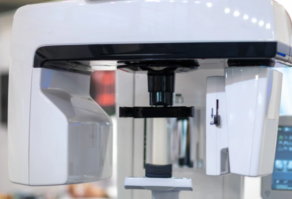
3D Cone Beam Imaging
3D dental imaging, or Cone Beam Computed Tomography (CBCT), offers a highly detailed view of your oral anatomy, providing clearer insights than traditional dental X-rays.
Understanding 3D CT Imaging
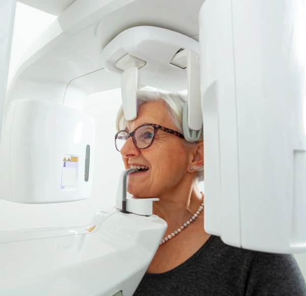
Traditional dental X-rays produce flat, two-dimensional images of small areas, offering only a limited view of your mouth. In contrast, 3D CT imaging, also called Cone Beam Computed Tomography (CBCT), provides a detailed, three-dimensional view of your entire oral and facial structure.
Think of it like the difference between looking at a single photo of a tooth versus rotating a 3D model to see every surface. CT imaging allows us to visualize not just individual teeth, but also your jaw, sinuses, and bone structures. This advanced technology enables more accurate diagnoses, precise treatment planning, and care that’s customized to your unique anatomy.
Why 3D CT Imaging Is Used
3D CT imaging with a Cone Beam Scanner improves every stage of diagnosis and treatment by providing a complete view of your oral structures. We may recommend a 3D scan in various situations to ensure safer, more precise, and more effective care, including:
- Diagnosing and planning treatment for TMJ (temporomandibular joint) disorders
- Designing precise surgical guides for dental implant placement
- Preparing for oral surgeries, including tooth extractions
- Checking root canal-treated teeth for potential infections along the roots
- Assessing airway volume to detect obstructions and evaluate sleep apnea
- Identifying bony defects or infections at previous extraction sites
How 3D CT Scanning Enhances Dental Care
Clear Visualization
Chairside monitors display detailed 3D images of your teeth and jaw, helping you easily understand problem areas and treatment options.
Better Communication
We can focus on specific areas, highlight concerns, and explain conditions with clarity and confidence.
Efficient Care Coordination
Digital files are easily shared with labs, insurance providers, or specialists, speeding up approvals and treatment timelines.
Ongoing Monitoring
3D scans are stored digitally, making it simple to compare future images, track progress, and adjust treatment plans as needed.